Saturday, November 17, 2012

Bibliography

Bibliography

Cook Rebecca, McFarland Kenneth. 2012. General Botany 111 Laboratory Manual. 14th Edition. 155-157 p.

Donner Josef. 1956. Rotifers. Stuttgart (Germany): W. Keller & Co. 5,65 p.

Gojdics Mary. 1953. Genus Euglena. Madison (WI): The University of Wisconson Press. 222 p.

Lee John J, Seymour Hunter H, Bovee Eugene C, editors. 1985. Illustrated Guide to Protozoa by Society of Protozoologists. Lawrence (KS): Allen Press, Inc. 200 p.

Patterson DJ. 2003. Free-Living Freshwater Protozoa. Washington DC: ASM Press. 17-18, 108, 125, 149 p.

Peachley Gordon. 1998. Pond I.  Blaine (WA): BioCam Communications Inc.

Pennak RW. 1989. Freshwater Invertebrates of the United States: Protozoa to Mollusca. 3rd Edition. New York: (NY)John Wiley and Sons. p 157, 164-165, 402-403.
 
Rainis Kenneth G., Russell Bruce J. 1996. Guide to Microlife. Canada: Franklin Watts.

Observation 4 - Nov. 13, 2012

This week, my microaquarium had far less activity than in the previous weeks.  Most of the organisms were in the bottom of the aquarium, and while they were slower moving, I could identify none that were dead; a lot of unidentified matter had collected at the bottom.  The numerous organisms identified last week as "planaria" were identified this week as spirostomia (not planaria) and they made up the bulk of the organisms found at the bottom of the tank.  Cyclidia and spirostomia are the most numerous organisms.  I saw no planaria this week, but did see amoebas, cyclidia, spirostomia, waterfleas, and the annelid "Chaetogaster" (Rainis and Russell) from last week.  One organism was visible to the naked eye, but not clearly enough to be identified.  The plants appear to have been eaten away, and only a few sprigs of both are left.  The aquarium was most active at the bottom, rather than the middle, as always before, and the overall movement of the organisms was slower.  No new organisms were identified.  It is my opinion that the aquarium will not be able to sustain itself much longer without additional nutrients and plants.
The above picture shows a portion of the body of the annelid "Chaetogaster."  I was unable to get a better picture because it was weaving in and out of the debris at the bottom of the tank.

Sunday, November 11, 2012

Week 3 Observation - November 6


Durling today's observation, I found that cyclidia, which commonly emerge in organically enriched cultures (Patterson 1996), remains the most abundant organism in the microaquarium, but the number of planaria have greatly increased, and this increase is what first caught my eye.  On Oct 23, I only saw two; last week I saw no increase in number; now there are between 20-30 traveling about all over the tank (but mainly in the middle) at a medium speed.  I recognized them, and their identification was confirmed by Dr. McFarland.  In the picture below is a planarium bent into a "J" shape.  Their presence implies good water quality.


With Dr. McFarland's help, I identified 4 new organisms in the microaquarium. 
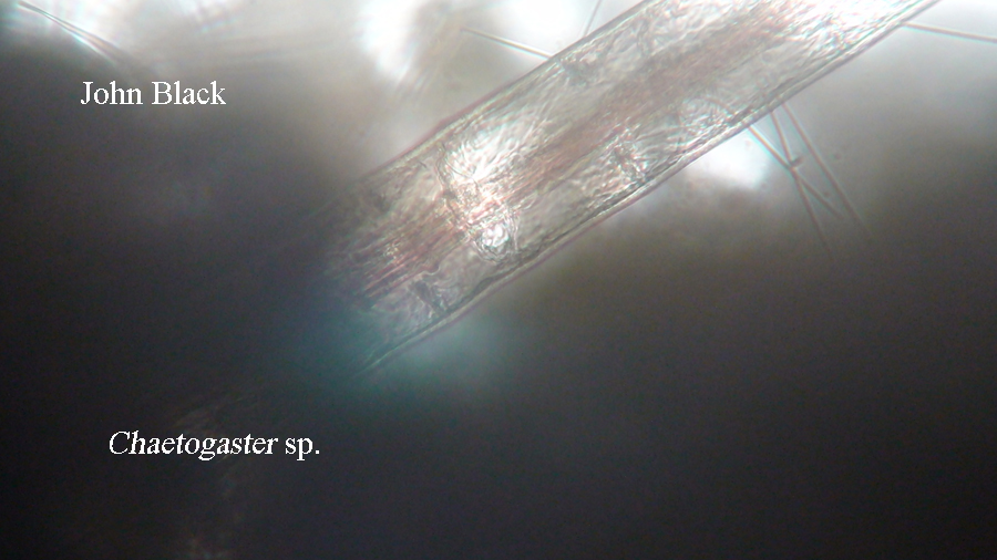
An annelid made its first appearance and was identified by Dr. McFarland.  I couldn't see its head, because it was hidden in the Amblestegium sp, but I could observe its segmented body.  It's the largest and "fiercest" looking organism I've seen in the microaquarium, and I only saw one.   


Also, the first appearance of a water flea, a type of gladocera, was noted (Pennak 1989).  It was eating a bit of something.


The third new organism was a euglena, E gracilis, which is a protist that has photosynthesizing chloroplasts (Gojdics 1953).  Its bright green color is seen in the picture below.  I only saw 2, and they were in the middle of the tank (a different area from the planaria) and completely still during observation.


The fourth new organism was a difflugia amoeba (Lee John J, Seymour Hunter H, Bovee Eugene C 1985).  I only saw one of these on the right, middle of the tank.  It moved a bit, but was not traveling around in the microaquarium.


Overall, the activity in the microaquarium has increased again this week.  With the exception of the increase in planaria, the numbers of other organisms have remained fairly consistent over the past 2 weeks.  I am identifying new organisms weekly.  The middle of the tank remains the most active area, and I identified no dead organisms.  I did add deionized water to refill the 0.5cm deficit of the microaquarium.
 
 
Bibliography
 
Cook Rebecca, McFarland Kenneth. 2012. General Botany 111 Laboratory Manual. 14th Edition. 155-157 p.
 
Donner Josef. 1956. Rotifers. Stuttgart (Germany): W. Keller & Co. 5,65 p.
 
Gojdics Mary. 1953. Genus Euglena. Madison (WI): The University of Wisconson Press. 222 p.
 
Lee John J, Seymour Hunter H, Bovee Eugene C, editors. 1985. Illustrated Guide to Protozoa by Society of Protozoologists. Lawrence (KS): Allen Press, Inc. 200 p.
 
Patterson DJ. 2003. Free-Living Freshwater Protozoa. Washington DC: ASM Press. 17-18, 108, 149 p.
 
Pennak RW. 1989. Freshwater Invertebrates of the United States: Protozoa to Mollusca. 3rd Edition. New York: (NY)John Wiley and Sons. p 157, 164-165, 402-403.


Sunday, November 4, 2012

Wk 2 Observation, Oct 30th

On Friday October 26th, one Beta Food Pellet was inserted into my Microaquarium. "Atison's Betta Food" is made by Ocean Nutrition, Aqua Pet Americas, 3528 West 500 South, Salt Lake City, UT 84104. The ingredients are: fish meal, wheat flower, soy meal, krill meal, minerals, vitamins and preservatives. The nutritional analysis is: crude protein 36%, crude fat 4.5%, crude fiber 3.5%, moisture 8%, and ash 15%.
Overall, the activity in the microaquarium continued to increase over that of the previous week.  Under the microscope, and with Dr. McFarland's assistance, I identified three new organisms and identified the "bubble-like" organisms seen in weeks one and two.

The first new organism identified was the amoeba shown in the picture below, which is the larger of the two I saw.  I observed movement inside the amoeba and was told by Dr. McFarland that the amoeba was digesting the food it had eaten.  The amoebas appeared to be stationary. 
The euchlanis rotifer, in the picture with the amoeba and also in the picture to the right, was identified last week.  They remain numerous and float about randomly at a medium speed.


Today was the first appearance of the philodina rotifer, seen in this picture above.  It was identified in the book Rotifers.  I saw only three of these, and each one was still except for the movement of it's mouth as it ate small particles floating in the water.

During the setup of the aquarium, and again last week, the organism in the picture below had remained too small to identify.  With Dr. McFarland's help, the "bubble-like" organism was identified as cyclidia.  These ciliates are the smallest, and remain the most numerous organism in the aquarium.  They moved in a jumping start/stop fashion and were rarely still.  The cilia are longer at one end than at the other.
 
 
A third new organism, Rotaria Neptuna, was identified with the help of Dr. McFarland and the book Rotifers.   Unfortunately, I am unable to download the video I made of this organism eating small particles.  The multicellular organism has a ring of cilia around its mouth, and its jaws looked to be spinning in circles as it ate; movement inside the organism is quite visible.  Then, for no apparent reason, it quickly and briefly withdrew, only to extend itself back into the picture and begin eating again.  I will make another attempt to download the video next week. 
 
I did see the organisms from last week, spirogyra, planaria, nematodes, tachyosoma, and the vorticella.  Their numbers remain basically unchanged.  There were many more filaments of oscillatoria present.
 
I saw no dead organisms, and the middle of the aquarium was the most active.